


A normal knee x-ray confirms that your knee joint is fundamentally sound by showing good alignment, healthy bone density, and adequate joint space. Knee X-rays are standard imaging procedures used to diagnose discomfort, oedema, damage, or arthritis. Understanding what constitutes a normal knee joint X-ray alleviates fears and enables appropriate next steps with confidence.
Normal Knee X-Ray
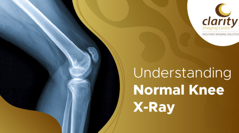
A normal knee x-ray shows smooth bone outlines, symmetrical joint spaces (3-5mm wide), and centred patella alignment in all standard knee x-ray views: anteroposterior (AP), lateral, and sunrise. Healthy knee x-ray results show no fractures, cartilage loss, or degenerative changes. Both the right knee x-ray and the normal left knee x-ray should show the same features; symmetry between the knees confirms normality.
Knee Anatomy on X-Ray
Radiology of the knee shows four major bones: the distal femur, the proximal tibia, the proximal fibula, and the patella. Key structures that are visible:
- Femoral Condyles: Rounded ends that form the medial and lateral joint surfaces.
- Tibial Plateau: The flat upper tibia bearing the weight of the femur
- Patella: The quadriceps tendon’s triangular sesamoid bone
- Joint Space: Cartilage appears as a radiolucent gap (3–5 mm).
- Fibula Head: Minimal articulation, lateral to the tibia
Normal lateral knee x-ray profiles show femur-tibia overlap, patellar location in the trochlear groove, and fabella (sesamoid variant).
Standard Knee X-Ray Views
Three projections provide a complete assessment:
- AP View: Joint space symmetry and alignment; front knee straight
- Lateral View: Patella position, posterior femoral condyles, side profile
- Sunrise/Axonometric: Trochlear tracking is evaluated using the Patella tangent
Abnormal knee x-ray contrasts include limited space (<3mm), osteophytes, or misalignment.
Findings of a Normal Knee X-Ray
Healthy knee x-ray characteristics include:
Bone Alignment and Contours
- Femoral condyles, smooth and symmetrical
- Tibial plateaus that run parallel to the condyles
- Absence of varus or valgus deformity
- Patella centred in the trochlear groove.
Joint Space
- Uniform bilateral breadth of 3-5 mm
- No constriction of the medial or lateral compartments
- Preserved cartilage thickness.
Bone Density & Texture
- Standard cortical outline (white borders)
- Uniform trabecular pattern
- Absence of erosions, cysts, or sclerosis
Soft Tissue Shadows
- A symmetrical suprapatellar pouch.
- Absence of joint effusion (dark pooling)
- Normal prepatellar soft tissue
Patellar Position
- Insall-Salvati ratio (normal height): 0.8–1.2
- Absence of subluxation or tilt
Also read: https://www.clarityimaging.in/bmd-test/
What Normal Results Mean for You
Seeing a normal knee x-ray usually reassures.
- No trauma-related fractures
- No osteoarthritis (preserved joint space)
- No problems with alignment that impact gait
- Strong bone health for bearing weight
Normal findings do not rule out soft-tissue issues (ligaments, meniscus), so an MRI is recommended when necessary. Pain with a standard X-ray frequently implies tendonitis, bursitis, or early cartilage concerns that can be managed conservatively.
Recognising Abnormal Knee X-Ray Findings
Contrast helps appreciation:
| Features | Normal Knee X-Ray | Abnormal Knee X-Ray |
| Joint Space | 3-5mm symmetrical | Narrowed (<2mm) |
| Bone Alignment | Neutral, Parallel | Varus/Valgus deformity |
| Patella | Centered | Tilted/Subluxed |
| Bone Texture | Smooth, Uniform | Osteophytes, Sclerosis |
| Soft Tissue | Even Shadows | Effusion, Calcification |
For high-quality image interpretation, get an X-ray test in Coimbatore from approved centers.
Age-Related Normal Variations
- Young Adults: smoother shapes and larger joint spaces
- Middle Age: Minimal medial narrowing is acceptable.
- Elderly: Slight 1-2mm narrowing without osteophytes is normal ageing.
Weight-bearing views are more sensitive to mild osteoarthritis than non-weight-bearing images.
When Knee X-Rays Are Ordered
Doctors frequently prescribe knee X-rays in cases of acute injuries from falls or sports, long-term pain and stiffness, or signs such as swelling, locking, or the knee giving way. They are also used to evaluate patients before arthroscopy and to monitor them after surgery. Clear or normal X-ray results might guide conservative care options, such as physiotherapy, injections, and lifestyle changes, before surgery.
Preparation & Procedure
The knee x-ray procedure takes 5-10 minutes:
- Remove clothing/jewellery above the knee.
- Sit or stand for positioning.
- Hold your breath for a moment.
- Minimal radiation (equal to a chest X-ray)
No special preparation is required. Usually, results are obtained the same day.
Next Steps After Normal Results
A typical knee X-ray eliminates doubt and offers clear paths to proactive well-being. Healthy findings often result in customised physical treatment focusing on quadriceps and hamstring training to stabilise the joint, as well as weight management through balanced eating and low-impact exercise (walking, swimming) to decrease mechanical stress.
If symptoms continue despite conservative therapy, consider advanced imaging, such as MRI, to check soft tissues (ligaments, meniscus) that are unseen on X-ray. This methodical strategy turns “normal” outcomes into empowered movement; to confidently monitor progress, speak with your orthopaedic physician or schedule dependable X-ray tests in Coimbatore.
This systematic technique transforms “normal” findings into empowered movement; see your orthopaedic doctor or receive dependable X-ray tests in Coimbatore follow-ups to track improvement with confidence.
Conclusion
A normal knee x-ray confirms structural integrity, removing concerns about fractures or severe arthritis. Smooth condyles, preserved joint space (3-5mm), and centred patella these characteristics indicate strong knees suited for daily use. Normal findings prioritise targeted rehabilitation above excessive worry. Consult an orthopaedist for personalised advice; healthy movement awaits.
FAQs
1. Difference between a normal and an abnormal knee X-ray lateral view?
A typical lateral view displays smooth joint surfaces and good alignment, but an aberrant view can reveal fractures, arthritic changes, or misalignment.
2. What is a normal knee X-ray AP lateral?
A normal AP and lateral knee X-ray reveals equal joint space, undamaged bones, and no evidence of oedema or degeneration.
3. How does a knee X-ray look?
The kneecap, joint space, shin bone, and thigh bone are all visible as a black-and-white image.
4. What is the anatomy of a knee X-ray?
It includes the femur, tibia, patella, fibula, joint space, and surrounding bone contours.
5. Is a knee X-ray safe?
Yes, it is generally safe and emits a small amount of radiation.
6. How long does a knee X-ray take?
The procedure usually takes about 5–10 minutes.
7. When is a knee X-ray recommended?
It is used to treat knee pain, damage, oedema, arthritis, and suspected fractures.


