


Quick Answer
A non-invasive cardiac ultrasound called a 2D echo test produces real-time moving images of the heart to evaluate its blood flow, shape, and function. Physicians use it to assess chamber size, valve performance, and ejection fraction. The test is safe for adults, elderly patients, and most pregnant women. It takes 20 to 45 minutes, usually doesn’t require fasting, and doesn’t include radiation.
Quick Overview Table
| Topic | Key Information |
| What is it? | Cardiac ultrasound imaging of heart structure and function in real time |
| Who needs it? | Patients with chest pain, breathlessness, palpitations, or high BP |
| What does it check? | Ejection fraction, valve function, chamber size, and fluid levels |
| Normal EF range? | Generally, 50% to 70%; always confirm with your cardiologist |
| Is it safe? | Yes, no radiation, non-invasive, safe for most patients |
| Approximate cost? | Roughly ₹800 to ₹3,500 in India; varies by city and centre type |
Table of Contents
- Introduction
- What Is a 2D Echo Test?
- Why Do Doctors Recommend a 2D Echo Test?
- Symptoms and Conditions Where a 2D Echo May Be Suggested
- 2D Echo Test Procedure Step by Step
- How to Prepare for a 2D Echo Test
- What Does a 2D Echo Test Report Show?
- 2D Echo Test Normal Report and Result Meaning
- 2D Echo Test Cost in India
- 2D Echo vs ECG vs Stress Test
- Benefits, Safety and Limitations of the 2D Echo Test
- When Should You Consult a Doctor After the Test?
- Conclusion
- FAQs
Introduction
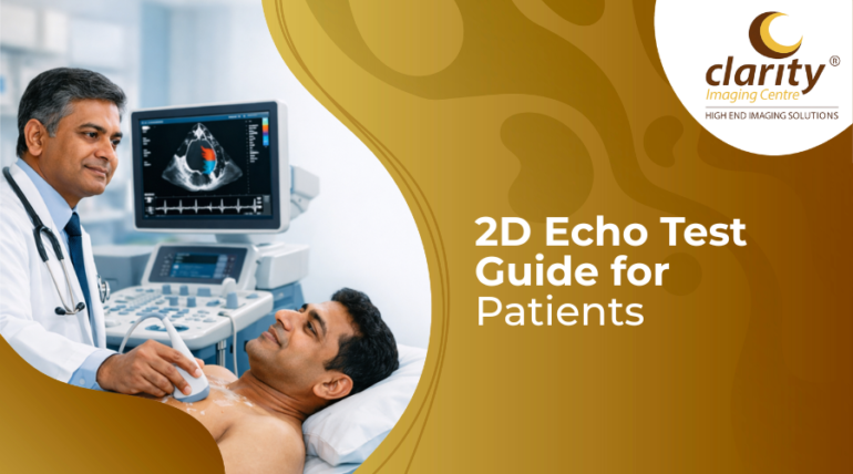
The 2D echo test is one of the most reliable methods physicians use to provide patients with information about heart health. This non-invasive cardiac imaging technique, which is short for two-dimensional echocardiography, uses ultrasonic waves to produce real-time, moving images of the heart, providing cardiologists with a comprehensive view of the heart’s appearance, motion, and function.
This blog describes everything from the test’s goal and process to the cost, report interpretation, and safety.
What Is a 2D Echo Test?
The 2D echo test, often known as the 2D echocardiography or simply the echo scan, is an ultrasound examination of the heart that provides two-dimensional cross-sectional images of the chambers, walls, and valves. These images move in real time, allowing the cardiologist to monitor the heart beating, accurately measure its size, and spot any structural or functional issues throughout the scan.
Why Do Doctors Recommend a 2D Echo Test?
A cardiologist or physician will recommend this test if they require a clear, thorough picture of how the heart is working. The test delivers information that cannot be obtained from an ECG or blood test alone.
Here are the main clinical reasons that clinicians recommend a 2D echo:
- Assess heart muscle function
- Evaluate heart valves
- Diagnose heart failure
- Detect pericardial effusion
- Pre- and post-surgery evaluation
- Monitor known heart conditions
Symptoms and Conditions Where 2D Echo May Be Suggested
The following situations usually result in a referral:
| Symptoms / Conditions | Why is Echo suggested? |
| Chest pain or tightness | Rules out structural cause; checks wall motion abnormalities |
| Shortness of breath | Assesses pumping efficiency and fluid around the heart |
| Heart palpitations | Evaluates chamber size and valve function behind irregular rhythm |
| High blood pressure | Checks for left ventricular hypertrophy from sustained pressure |
| Diabetes (long-standing) | Screens for diabetic cardiomyopathy affecting the heart muscle |
| Heart murmur detected | Identifies valve disease behind the abnormal heart sound |
| Prior heart attack | Measures post-infarct ejection fraction and wall damage |
| Pre-operative clearance | Confirms cardiac fitness before a major surgical procedure |
2D Echo Test Procedure Step by Step
The 2D echo test procedure is painless, non-invasive, and typically takes 20 to 45 minutes. Here’s what to expect at each stage of your appointment:
- Step 1 – Preparation and positioning
- Step 2 – Left-side positioning
- Step 3 – Gel application
- Step 4 – Transducer movement
- Step 5 – Image and measurement capture
- Step 6 – Doppler assessment (if required)
- Step 7 – Report generation
How to Prepare for a 2D Echo Test?
A standard resting echocardiogram does not need fasting. You can eat, drink, and take your regular medications before the test, unless your doctor gives you alternative instructions, like the following:
- Clothing
- Medicines
- Avoid lotions on the chest
- Special instructions apply for
What Does a 2D Echo Test Report Show?
Understanding the important parameters allows you to have a better-informed conversation with your cardiologist following the scan:
| Report Parameter | What It Tells the Doctor |
| Ejection Fraction (EF) | The percentage of blood pumped per heartbeat reflects pump efficiency |
| Left Ventricular Size | Chamber dimensions: enlarged size may indicate cardiomyopathy |
| Wall Thickness | Detects hypertrophy from high BP or athletic conditioning |
| Diastolic Function | How well the heart relaxes between beats affects filling efficiency |
| Pericardial Effusion | Presence and volume of fluid in the sac surrounding the heart |
| Right Heart Parameters | Pulmonary pressures and right ventricular function assessment |
2D Echo Test Normal Report and Result Meaning
A normal 2D echo test result typically includes an ejection fraction between 50% and 70%, normal valve appearance and movement, no considerable pericardial effusion, and chamber dimensions that are within age-appropriate ranges. These findings, considered collectively, indicate that the heart is physically and physiologically healthy.
However, a single metric significantly beyond the reference range does not automatically indicate illness. Report interpretation must always take into account the patient’s age, physical size, symptoms, and clinical history.
2D Echo Test Cost in India
Government hospital rates are often lower than those of commercial diagnostic chains or specialist cardiac facilities.
| Scan Type | Approximate Price Range (India) |
| Standard 2D Echo | ₹800 – ₹1,800 |
| 2D Echo with Colour Doppler | ₹1,200 – ₹2,500 |
| Stress Echocardiography | ₹2,500 – ₹6,000 |
| Transoesophageal Echo (TEE) | ₹4,000 – ₹8,000 |
| Corporate / Insurance covered | Varies; confirm with your provider before booking |
2D Echo vs ECG vs Stress Test
Here’s a straightforward comparison to help you understand what each test does and when it’s usually recommended:
| Feature | 2D Echo Test | ECG | Stress Test |
| What it shows | Heart structure and function | Electrical rhythm of the heart | Heart performance under exertion |
| Technology used | Ultrasound (no radiation) | Electrical impulse recording | ECG + exercise or medication |
| Duration | 20–45 minutes | 5–10 minutes | 30–60 minutes |
| Detects blockage? | Indirectly (wall motion) | Partially (ischaemia signs) | More specifically than resting ECG |
| Fasting required? | Usually not | No | Yes, typically 3–4 hours |
| Best for | Valve, pump and structural | Rhythm and conduction issues | Exertional chest pain, fitness |
Benefits, Safety and Limitations of 2D Echo Test
The 2D echo test does not employ ionising radiation; hence, it is perfectly safe for repeated use. It delivers real-time dynamic images, which static examinations such as X-rays cannot provide. It is readily available, reasonably priced, and can be performed at the bedside for severely ill patients as needed.
- Non-invasive
- No radiation
- Real-time imaging
- Limitation – image quality
- Limitation – coronary arteries
A reputable ultrasound scan centre in Coimbatore that provides obstetric and cardiac imaging services can coordinate both maternal and foetal echocardiograms in a single consultation if clinically acceptable.
When Should You Consult a Doctor After the Test?
Do not delay a consultation if your report contains any of the following:
- Ejection fraction below 40%
- Moderate or severe valve disease
- Pericardial effusion of significant volume
- Wall motion abnormalities
- Elevated estimated pulmonary pressure
Disclaimer
The information in this article is meant solely for educational awareness and general information. It is not intended to replace professional medical advice, diagnosis, or treatment. Always consult a qualified healthcare professional or doctor regarding any medical condition or health concern.
Conclusion
A 2D echo test is one of the most useful, accessible, and patient-friendly cardiac diagnostic techniques available today. It provides cardiologists with a real-time view of how the heart is performing, aids in the early detection of abnormalities, and guides proper therapy all without the need for radiation, pain, or long preparation.
Concerned about your heart health?
Book your 2D Echo Test today and get expert guidance with a safe, painless heart scan.
FAQs
1. What is a Carotid Doppler test?
A Carotid Doppler test is an ultrasound examination that measures the flow of blood via the neck’s carotid arteries.
2. Why is a Carotid Doppler ultrasound done?
Carotid Doppler ultrasound is used to detect stenosis, blockages, or diminished blood flow in the carotid arteries.
3. Is a Carotid Doppler test painful?
No, a Carotid Doppler test is painless, non-invasive, and does not require radiation.
4. What are normal values in a Carotid Doppler report?
Normal readings suggest smooth blood flow in the carotid arteries, with no major narrowing or obstruction.
5. How long does a Carotid Doppler ultrasound take?
Carotid Doppler ultrasounds normally take 15 to 30 minutes to complete.
6. Can Carotid Doppler detect stroke risk?
Ear bone injury can cause hearing loss, ear pain, dizziness, ringing in the ears, and facial weakness.
7. What happens if the Carotid Doppler result is abnormal?
If the results are abnormal, your doctor may recommend medications, lifestyle changes, additional tests, or treatment to improve blood flow.
8.What is the cost of a Carotid Doppler test in India?
Carotid Doppler tests in India typically cost between ₹1,500 and ₹4,000, based on hospital and location.


